A fungus that looks like shooting stars. A dog’s eye that looks like the moon over a lake. And a fish that resembles a Native American headdress.
Cross sections of biological specimens — “images the naked eye can’t see” — adorn the lobby of the The Wistar Institute at 36th and Spruce streets as part of a Nikon-sponsored photomicrography exhibition. Wistar, an independent medical research facility on Penn’s campus, has hosted the Nikon Small World Competition several times in the past.
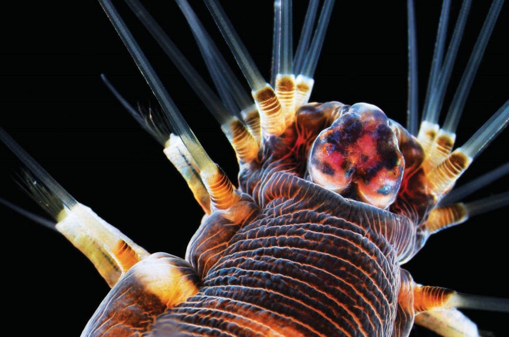
Photomicrography involves taking pictures of magnified objects through a microscope. The annual international contest encourages anyone with a microscope to submit an image to the Nikon website.
The competition started in 1977, and winners are selected by a panel of judges that this year included a National Geographic photojournalist and biomedical researchers. The images will be on display at Wistar until March 7.
The Wistar Institute’s Microscopy Core Facility Manager James Hayden has placed in the top 20 in the competition 13 times and judged the competition once. In 1999, his photos took the third, sixth and eighth place awards.
“My particular technique looks for things that look like something else,” he said. “So you look at it and it’s not so much, ‘Oh, that’s a neuron,’ instead it’s more like, ‘Oh, what is that?’”
He emphasized the importance of visual impact, uniqueness and technical proficiency in the judging process. Many of the images in the top 20 use a technique called “image stacking,” which consists of taking multiple photographs in order to add more detail and depth to the image.
Hayden believes that photomicrography does have applications in scientific research, but the photos on display are tailored artistically. The intention is to draw attention rather than capturing all the details of an organism.
The first-place image is a spiral-shaped colonial plankton with detailed grooves. The second-place image is a close-up of a turtle’s retina but almost resembles red and orange Skittles on a light blue background.
The first-place photographer and curator of the online Micropolitan Museum, Wim van Egmond, does not work in a lab, unlike many of the other participants. Van Egmond, who is from The Netherlands, has participated in the competition for many years.
Hayden thinks that photomicrography lies at the intersection of art and science. “The more you experiment with science, the more you come up with these views that people have never seen,” he said.
