Researchers at the Hospital of the University of Pennsylvania published a report that could help bring new technology to the forefront of breast cancer screening.
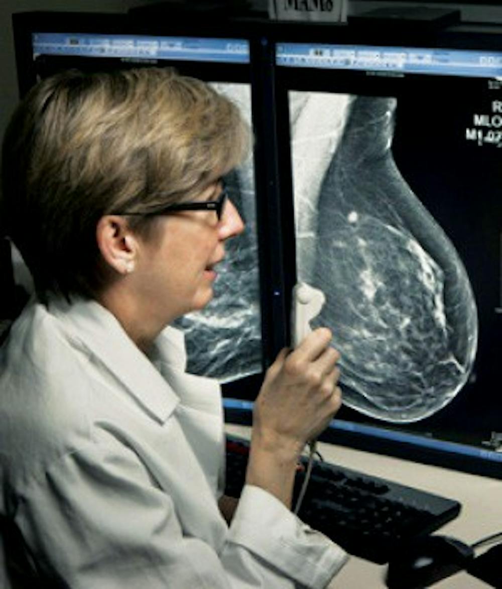
Tomosynthesis, or 3D mammography, which was approved by the Food and Drug Administration in 2011, created a 3D reconstruction of breast tissue and allows radiologists to see the overlapping tissue more clearly.
The study, published in the June 25 issue of Journal of the American Medical Association, was conducted on about half a million women — the largest study yet to measure on the effectiveness of tomosynthesis.
According to HUP Chief of Breast Imaging and senior author of the paper Dr. Emily Conant, tomosynthesis showed significant increase in detection rates and reduced recall rates in all patients, a finding that Conant and her team had not expected.
“We expected that 3D would reduce false positive exams, which it did,” she said. “We did not expect that it would increase cancer detection as much as it did, and were particularly happy to see that increased detection represented invasive cancers, which are important to detect.”
Compared to screening that only used traditional digital mammography, detection rates rose by 41 percent and recall rates fell by 15 percent when tomosynthesis was coupled with digital mammography.
Conant and her fellow researchers have been studying this innovative technology for about ten years. When the FDA approved a clinical unit for patient use in 2011, the researchers began using tomosynthesis with all patients who came in for routine mammograms.
“Technology is not a static thing. It’s an evolving platform,” Conant said, in reference to the future use of tomosynthesis.
“It’s continued to evolve and improve; we’ve now brought the x-ray dose down with the new technique because we have an ability to create reconstructed 2D images to accompany the 3D images.”
Conant eluded to the future of her team’s tomosythesis research as well. Future research will study further improvements of the technology and get deeper into patient-level data.
Staff Writer Alison Elliot contributed reporting.
